Details
Structure visualisation
Entry information
| Complex | |
| AACDB_ID: | 6708 |
| PDBID: | 3V4V |
| Chains: | HL_B |
| Organism: | Homo sapiens, Mus musculus |
| Method: | XRD |
| Resolution (Å): | 3.10 |
| Reference: | 10.1083/jcb.201110023 |
| Antibody | |
| Antibody: | Act-1 Fab |
| Antibody mutation: | No |
| INN (Clinical Trial): | |
| Antigen | |
| Antigen: | Integrin beta-7 |
| Antigen mutation: | No |
| Durg Target: | P26010 |
Sequence information
Antibody
Heavy Chain: H
Mutation: NULL
| >3V4V_H|Chain C[auth H], G[auth M]|MONOCLONAL ANTIBODY Act-1 HEAVY CHAIN|Mus musculus (10090) QVQLQQPGAELVKPGTSVKLSCKGYGYTFTSYWMHWVKQRPGQGLEWIGEIDPSESNTNYNQKFKGKATLTVDISSSTAYMQLSSLTSEDSAVYYCARGGYDGWDYAIDYWGQGTSVTVSSAKTTPPSVYPLAPGSAAQTNSMVTLGCLVKGYFPEPVTVTWNSGSLSSGVHTFPAVLESDLYTLSSSVTVPSSPRPSETVTCNVAHPASSTKVDKKIV |
Light Chain: L
Mutation: NULL
| >3V4V_L|Chain D[auth L], H[auth N]|MONOCLONAL ANTIBODY Act-1 LIGHT CHAIN|Mus musculus (10090) DVVVTQTPLSLPVSFGDQVSISCRSSQSLAKSYGNTYLSWYLHKPGQSPQLLIYGISNRFSGVPDRFSGSGSGTDFTLKISTIKPEDLGMYYCLQGTHQPYTFGGGTKLEIKRADAAPTVSIFPPSSEQLTSGGASVVCFLNNFYPKDINVKWNIDGSERQNGVLNSWTDQDSKDSTYSMSSTLTLTKDEYERHNSYTCEATHKTSTSPIVKSFNRN |
Antigen
Chain: B
Mutation: NULL
| >3V4V_B|Chain B, F[auth D]|Integrin beta-7|Homo sapiens (9606) ELDAKIPSTGDATEWRNPHLSMLGSCQPAPSCQKCILSHPSCAWCKQLNFTASGEAEARRCARREELLARGCPLEELEEPRGQQEVLQDQPLSQGARGEGATQLAPQRVRVTLRPGEPQQLQVRFLRAEGYPVDLYYLMDLSYSMKDDLERVRQLGHALLVRLQEVTHSVRIGFGSFVDKTVLPFVSTVPSKLRHPCPTRLERCQSPFSFHHVLSLTGDAQAFEREVGRQSVSGNLDSPEGGFDAILQAALCQEQIGWRNVSRLLVFTSDDTFHTAGDGKLGGIFMPSDGHCHLDSNGLYSRSTEFDYPSVGQVAQALSAANIQPIFAVTSAALPVYQELSKLIPKSAVGELSEDSSNVVQLIMDAYNSLSSTVTLEHSSLPPGVHISYESQCEGPEKREGKAEDRGQCNHVRINQTVTFWVSLQATHCLPEPHLLRLRALGFSEELIVELHTLCDCNCSDTQPQAPHCSDGQGHLQCGVCSCAPGRLGRLCESRGLENLYFQ |
Interaction
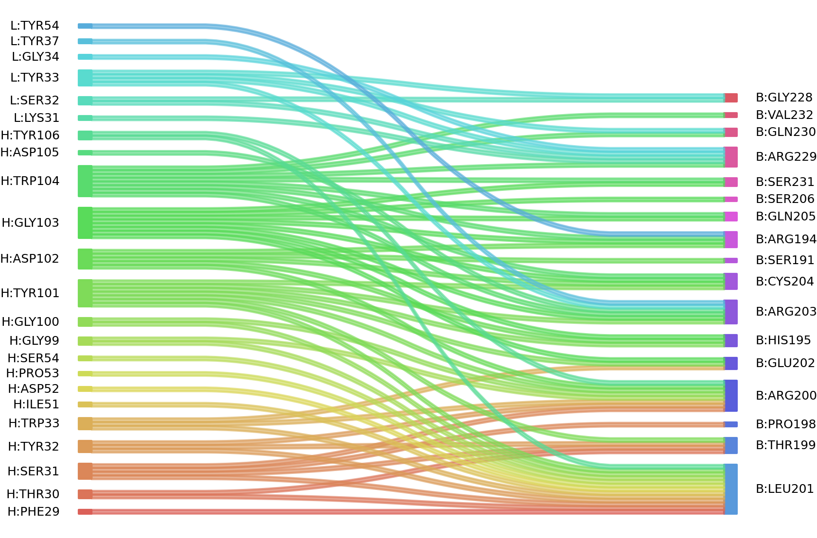
1、Solvent accessible surface areas (SASA) were calculated (Naccess V2.1.1) for each residue in antibody and antigen, respectively. The residues with SASA loss in binding of more than 1Å2 were classified as interacting residues.
Interacting residues (ΔSASA based)
| Chain residues position delta_SASA
: residuesposition H: THR30 SER31 TYR32 TRP33 ASP52 PRO53 SER54 GLY100 TYR101 ASP102 GLY103 TRP104 L: SER32 TYR33 GLY34 TYR54 B: ARG194 HIS195 PRO198 THR199 ARG200 LEU201 GLU202 ARG203 CYS204 GLN205 GLY228 ARG229 GLN230 SER231 |
2、We defined interacting paratope-epitope residues by a distance cutoff of < 6 Å . Two amino acids are considered as interacting residues if they have at least one atom within a distance of 6 Å from any atom.
Interacting residues (Atom distance based)
